
BY CLIVE LATHEY, D.O MSC (SPORTS MEDICINE)
Musculoskeletal pain (back, neck and other joints), can be very acute and debilitating; resulting in fear and anxiety. As a consequence, patients often request MRI scans in the belief that it is going to be an important part of their treatment. While MRI scans are a “gold standard” diagnostic investigation, there are guidelines concerning their appropriate use.
History of MRI scans
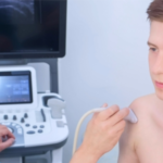
MRI scans were developed in the 1970s and 1980s as a medical imaging technique that did not use radiation. Instead they work using a strong magnetic field and radio waves to generate images of the body. MRI scans essentially map the location of water and fat and as most body structures contain both, an image is created. The physics involved in achieving this is complicated, but the end result has helped revolutionise medical investigation.
MRI scans have a wide range of application from investigation of the brain and spine to other joints and organs, including the heart and liver. Although MRI machines are expensive, costing up to £1 million pounds, there are estimated to be around 35,000 in use worldwide and approximately 497 in the UK. Despite having fewer MRI scans than most western nations, the cost of an MRI scan in the UK can be as little as £260 in specialist diagnostic centres and as much as £800 – £1,000 in private hospitals.
Appropriate use
X-rays are generally used to investigate bone pathology, such as fractures, while MRI scans are used to examine soft tissues such as muscles, ligaments, cartilage, blood vessels, nerves and discs. MRI scans are often recommended for any of the following:
- Unexplained pain
- Accidents and trauma
- Acute nerve pain, such as Sciatica
- When conservative treatment (such as physical therapy) is not helping
- Acute pain lasting more than 4-6 weeks, which is not improving
- Neurological symptoms
- Suspected disc prolapse in the spine
- Other “red flag” signs
Most acute back and neck pain recovers within 4-6 weeks with appropriate management, advice and exercises provided by a physiotherapist or an osteopath.
The guidelines are, in essence:
- Use non-steroidal anti-inflammatory medication (NSAIDs)
- Refer to physical therapy/physiotherapy
- Refrain from early MRI referral, apart from exceptional cases (red flags)
- Avoid habit forming medication
- Avoid early referral for injections or surgery
Some surprising facts
MRI scans reveal considerable detail about our internal bodies. The problem is that very often so called abnormalities do not in fact not cause pain. Osteoarthritis (normal wear and tear) in our joints and bones may look bad, but it may not be causing symptoms. For example, MRI scans of the following:
- Disc bulging occurs in 87% of people aged between 20-70 years
- Disc degeneration: 37% (20 year olds), 96% (80 year olds)
- Hips: 2,114 people studied; 67% had arthritis and no pain
- Knees: 5,397 people studied; 43% had arthritis and no pain.
- Shoulder: 40-70 year age group. 78% had a pathology and no pain.
It is therefore very important that a patient is physically examined and a detailed case history is taken in conjunction with an MRI investigation. It is only when you have all the clinical information and the MRI results that a diagnosis can be made. While MRI scans are the “gold standard” investigation it is important to remember that people can adapt to conditions like arthritis and be relatively pain free, particularly if they have good levels of fitness.
The “Nocebo” effect
If a medical professional advises an MRI scan and the diagnosis is “degenerative disc disease”, you now have a label. In fact, you have just been told you have a disease. This feeds anxiety and fear creating a “nocebo” effect (the opposite of a placebo effect). This term applies to situations where a diagnosis can create a negative state of mind, which has an adverse effect on health.
This poses wider reaching questions. What are the boundaries that define an illness or pathology? What is, in fact, normal? If the data tells us that 98% of people with acute lower-back pain (along with structural changes) on MRI have just suffered a sprain, we clearly need a more careful interpretation of these results. Definitively, lower-back pain should never be diagnosed from a scan alone.
Over-diagnosis in back pain
Over-diagnosis as a concept may sound counter-intuitive and, indeed, many people’s lives have benefited from appropriate use of MRI scans. However, this final statistic delivers perhaps the strongest message. In the United States, people referred for MRI scans for routine back pain are eight times more likely to have surgery. Now, that is a statistic that is definitely worth changing. Here is a great link to a podcast on the subject of over-diagnosis by Ray Moynihan.
Further viewing
Below is an interesting video on the subject by Doctor Bahram Jam, founder and director of Advanced Physical Therapy Education Institute (APTEI).
Appointments
If you are experiencing back or neck pain, make an appointment to see one of our osteopaths or physiotherapists. We will take a detailed history followed by a thorough examination and clinical tests. We will organise MRI scans and other investigations if it is appropriate.
You can book online or call us on 0208 789 3881.